Posted on

The Vikings Were Advanced Dentists

This image has an empty alt attribute; its file name is ai-free.png

Widespread caries and toothache – but also some dental work and filing of front teeth. Viking Age teeth from Varnhem in Sweden bear witness to surprisingly advanced dentistry. This has been shown in a study carried out at the University of Gothenburg.

The study examined 3,293 teeth from 171 individuals among the Viking Age population. The site is known for extensive excavations of Viking and medieval environments, including tombs where skeletons and teeth have been preserved well in favorable soil conditions.
The teeth underwent clinical examinations using standard dentistry tools under bright light. A number of X-ray examinations were also performed using the same technique used in dentistry, where the patient bites down on a small square imaging plate in the mouth.
The results, which have been published in the journal PLOS ONE, show that 49% of the Viking population had one or more caries lesions. Of the adults’ teeth, 13% were affected by caries – often at the roots. However, children with milk teeth – or with both milk and adult teeth – were entirely caries-free (https://journals.plos.org/plosone/article?id=10.1371/journal.pone.0295282).
The findings suggest that caries, tooth infections, and toothache were common among the Viking population in Varnhem. However, the study also reveals examples of attempts to look after teeth in various ways.
“There were several signs that the Vikings had modified their teeth, including evidence of using toothpicks, filing front teeth, and even dental treatment of teeth with infections,” says Carolina Bertilsson, a dentist and Associate Researcher, and the study’s first-named and corresponding author.
One sign of more sophisticated procedures was molars with filed holes, from the crown of the tooth and into the pulp, probably in order to relieve pressure and alleviate severe toothache due to infection.
The filed front teeth may have been a form of identity marker. In both this and previous studies, the cases found were male. Carolina Bertilsson continues:
“This study provides new insights into Viking oral health, and indicates that teeth were important in Varnhem’s Viking culture. It also suggests that dentistry in the Viking Age was probably more sophisticated than previously thought.”

AR #84

The New Crystal Skull Enigma

by Phillip Coppens

Posted on

Brains Can Now Communicate with Thought Alone

This image has an empty alt attribute; its file name is ai-free.png

A speech prosthetic developed by a collaborative team of Duke neuroscientists, neurosurgeons, and engineers can translate a person’s brain signals into what they’re trying to say.

Appearing in the journal Nature Communications, the new technology might one day help people unable to talk due to neurological disorders regain the ability to communicate through a brain-computer interface.
“There are many patients who suffer from debilitating motor disorders, like ALS (amyotrophic lateral sclerosis) or locked-in syndrome, that can impair their ability to speak,” said Gregory Cogan, PhD, a professor of neurology and neurosurgery at Duke and one of the lead researchers involved in the project. “But the current tools available to allow them to communicate are generally very slow and cumbersome.”
Imagine listening to an audiobook at half-speed. That’s the best speech decoding rate currently available, which clocks in at about 78 words per minute. People, however, speak around 150 words per minute.
The lag between spoken and decoded speech rates is partially due the relatively few brain activity sensors that can be fused onto a paper-thin piece of material that lays atop the surface of the brain. Fewer sensors provide less decipherable information to decode.
To improve on past limitations, Cogan teamed up with Jonathan Viventi, PhD, associate professor of biomedical engineering, whose lab specializes in making high-density, ultra-thin, and flexible brain sensors.
Viventi and his team packed a record-breaking 256 microscopic brain sensors onto a postage stamp-sized piece of flexible, medical-grade plastic. Neurons just a grain of sand apart can have wildly different activity patterns when coordinating speech, so it’s necessary to distinguish signals from neighboring brain cells to help make accurate predictions about intended speech.
After fabricating the new implant, Cogan and Viventi teamed up with several Duke University Hospital neurosurgeons, including Derek Southwell, MD, PhD, Nandan Lad, MD, PhD, and Allan Friedman, MD, who helped recruit four patients to test the implants. The experiment required the researchers to place the device temporarily in patients who were undergoing brain surgery for some other condition, such as  treating Parkinson’s disease or having a tumor removed. Time was limited for Cogan and his team to test drive their device in the OR.
Read the full story from Duke Institute of Brain Sciences on Duke Today and in Newsweek.

AR #79

Brain to Brain Communication

Posted on

Warm Feelings from Missing Hands

This image has an empty alt attribute; its file name is ai-free.png

An unexpected discovery about temperature feedback has led to new bionic technology that allows amputees to sense the temperature of objects—both hot and cold—directly in the phantom hand. The technology opens up new avenues for non-invasive prosthetics.

“When I touch the stump with my hand, I feel tingling in my missing hand, my phantom hand. But feeling the temperature variation is a different thing, something important… something beautiful,” says Francesca Rossi.

Rossi is an amputee from Bologna, Italy. She recently participated in a study to test the effects of temperature feedback directly to the skin on her residual arm. She is one of 17 patients to have felt her phantom, missing hand, change in temperature thanks to new EPFL technology. More importantly, she reports feeling reconnected to her missing hand.

“Temperature feedback is a nice sensation because you feel the limb, the phantom limb, entirely. It does not feel phantom anymore because your limb is back,” Rossi continues.

Researchers Silvestro Micera and Solaiman Shokur have been keen on incorporating new sensory feedback into prosthetic limbs for providing more realistic touch to amputees, and their latest study focuses on temperature. They stumbled upon a discovery about temperature feedback that far exceeds their expectations.
If you place something hot or cold on the forearm of an intact individual, that person will feel the object’s temperature locally, directly on their forearm. But in amputees, that temperature sensation on the residual arm may be felt­… in the phantom, missing hand.

By providing temperature feedback non-invasively, via thermal electrodes (aka thermodes) placed against the skin on the residual arm, amputees like Rossi report feeling temperature in their phantom limb. They can feel if an object is hot or cold, and can tell if they are touching copper, plastic or glass. In a collaboration between EPFL, Sant’Anna School of Advanced Studies (SSSA) and Centro Protesi Inail, the technology was successfully tested in 17 out of 27 patients. The results are published in Science. (https://ieeexplore.ieee.org/abstract/document/6775436)
“Of particular importance is that phantom thermal sensations are perceived by the patient as similar to the thermal sensations experienced by their intact hand,” explains Shokur, EPFL senior scientist neuroengineer who co-led the study.

The projection of temperature sensations into the phantom limb has led to the development of new bionic technology, one that equips prosthetics with non-invasive temperature feedback that allows amputees to discern what they’re touching.

“Temperature feedback is essential for relaying information that goes beyond touch, it leads to feelings of affection. We are social beings and warmth is an important part of that,” says Micera, Bertarelli Foundation Chair in Translational Neuroengineering, professor at EPFL and SSSA who also co-led the study. “For the first time, after many years of research in my laboratory showing that touch and position information can be successfully delivered, we envisage the possibility of restoring all of the rich sensations that one’s natural hand can provide.”

AR #90

The Paraffin Mold Experiments

By Michael Tymn

Posted on

Could Microscopic Medical Robots Travel Through Your Body?

By Daniel Strain

This image has an empty alt attribute; its file name is ai-free.png

A team of engineers at the CU Boulder has designed a new class of tiny, self-propelled robots that can zip through liquid at incredible speeds—and may one day even deliver prescription drugs to hard-to-reach places inside the human body.

The researchers describe their mini healthcare providers in a paper published last month in the journal Small.

“Imagine if microrobots could perform certain tasks in the body, such as non-invasive surgeries,” said Jin Lee, lead author of the study and a postdoctoral researcher in the Department of Chemical and Biological Engineering. “Instead of cutting into the patient, we can simply introduce the robots to the body through a pill or an injection, and they would perform the procedure themselves.”

Lee and his colleagues aren’t there yet, but the new research is big step forward for tiny robots. 
The group’s microrobots are really small. Each one measures only 20 micrometers wide, several times smaller than the width of a human hair. They’re also really fast, capable of traveling at speeds of about 3 millimeters per second, or roughly 9,000 times their own length per minute. That’s many times faster than a cheetah in relative terms. 

They have a lot of potential, too. In the new study, the group deployed fleets of these machines to transport doses of dexamethasone, a common steroid medication, to the bladders of lab mice. The results suggest that microrobots may be a useful tool for treating bladder diseases and other illnesses in people.

“Microscale robots have garnered a lot of excitement in scientific circles, but what makes them interesting to us is that we can design them to perform useful tasks in the body,” said C. Wyatt Shields, a co-author of the new study and assistant professor of chemical and biological engineering.

If that sounds like something ripped from science fiction, that’s because it is. In the classic film Fantastic Voyage, a group of adventurers travels via a shrunken-down submarine into the body of a man in a coma.“The movie was released in 1966. Today, we are living in an era of micrometer- and nanometer-scale robots,” Lee said.

He imagines that, just like in the movie, microrobots could swirl through a person’s blood stream, seeking out targeted areas to treat for various ailments.

The team makes its microrobots out of materials called biocompatible polymers using a technology similar to 3D printing. The machines look a bit like small rockets and come complete with three tiny fins. They also include a little something extra: Each of the robots carries a small bubble of trapped air, similar to what happens when you dunk a glass upside-down in water. If you expose the machines to an acoustic field, like the kind used in ultrasound, the bubbles will begin to vibrate wildly, pushing water away and shooting the robots forward. 

AR #84

Molecular Machines that Defy Darwin

by Casey Luskin

Posted on

Do ‘Stuck’ Stem Cells Account for Gray Hair?

Certain stem cells have a unique ability to move between growth compartments in hair follicles, but get stuck as people age and so lose their ability to mature and maintain hair color, a new study shows.

Led by researchers from NYU Grossman School of Medicine, the new work focused on cells in the skin of mice and also found in humans called melanocyte stem cells, or McSCs. Hair color is controlled by whether nonfunctional but continually multiplying pools of McSCs within hair follicles get the signal to become mature cells that make the protein pigments responsible for color.

Published in the journal Nature online April 19, the new study showed that McSCs are remarkably plastic. This means that during normal hair growth, such cells continually move back and forth on the maturity axis as they transit between compartments of the developing hair follicle. It is inside these compartments where McSCs are exposed to different levels of maturity-influencing protein signals.

Specifically, the research team found that McSCs transform between their most primitive stem cell state and the next stage of their maturation, the transit-amplifying state, and depending on their location.

The researchers found that as hair ages, sheds, and then repeatedly grows back, increasing numbers of McSCs get stuck in the stem cell compartment called the hair follicle bulge. There, they remain, do not mature into the transit-amplifying state, and do not travel back to their original location in the germ compartment, where WNT proteins would have prodded them to regenerate into pigment cells.

AR #74

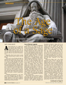
The Age of a Sage

by John White

Posted on

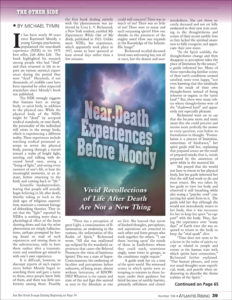
Conscious-Like Activity Found In the Dying Brain

Reports of near-death experiences—with tales of white light, visits from departed loved ones, hearing voices, among other attributes—capture our imagination and are deeply engrained in our cultural landscape.

The fact that these reports share so many common elements begs the question of whether there is something fundamentally real underpinning them—and that those who have managed to survive death are providing glimpses of a consciousness that does not completely disappear, even after the heart stops beating.

A study published in the Proceedings of the National Academy of Science, provides early evidence of a surge of activity correlated with consciousness in the dying brain.

The study, led by Jimo Borjigin, Ph.D., associate professor in the Department of Molecular & Integrative Physiology and the Department of Neurology, and her team is a follow up to animal studies conducted almost 10 years ago in collaboration with George Mashour, M.D., Ph.D., the founding director of the Michigan Center for Consciousness Science.

Similar signatures of gamma activation were recorded in the dying brains of both animals and humans upon a loss of oxygen following cardiac arrest.

“How vivid experience can emerge from a dysfunctional brain during the process of dying is a neuroscientific paradox. Dr. Borjigin has led an important study that helps shed light on the underlying neurophysiologic mechanisms,” said Mashour.

The team identified four patients who passed away due to cardiac arrest in the hospital while under EEG monitoring. All four of the patients were comatose and unresponsive. They were ultimately determined to be beyond medical help and, with their families’ permission, removed from life support.

Upon removal of ventilator support, two of the patients showed an increase in heart rate along with a surge of gamma wave activity, considered the fastest brain activity and associated with consciousness.

Furthermore, the activity was detected in the so-called hot zone of neural correlates of consciousness in the brain, the junction between the temporal, parietal and occipital lobes in the back of the brain. This area has been correlated with dreaming, visual hallucinations in epilepsy, and altered states of consciousness in other brain studies.

These two patients had previous reports of seizures, but no seizures during the hour before their deaths, explains Nusha Mihaylova, M.D., Ph.D., a clinical associate professor in the Department of Neurology who has collaborated with Borjigin since 2015 by collecting EEG data from deceased patients under intensive care unit treatment. The other two patients did not display the same increase in heart-rate upon removal from life support, nor did they have increased gamma activity.

Because of the small sample size, the authors caution against making any global statements about the implications of the findings. They also note that it’s impossible to know in this study what the patients experienced because they did not survive.

“We are unable to make correlations of the observed neural signatures of consciousness with a corresponding experience in the same patients in this study. However, the observed findings are definitely exciting and provide a new framework for our understanding of covert consciousness in the dying humans,” she said.

Larger, multi-center studies including EEG-monitored ICU patients who survive cardiac arrest, could provide much needed data to determine whether or not these bursts in gamma activity are evidence of hidden consciousness even near death.

Additional authors on this paper include Gang Xu, Duan Li, Fangyun Tian, Peter M. Farrehi, Jack M. Parent and Michael Wang Paper cited: “Surge of neurophysiological coupling and connectivity of gamma oscillations in the dying human brain,” Proceedings of the National Academy of Science. https://www.pnas.org/doi/10.1073/pnas.2216268120

AR #106

Near Death Experience Before Moody

by Michael Tymn

Posted on

Does Newly Discovered Intracellular Electricity Power Biology?

The human body relies heavily on electrical charges. Lightning-like pulses of energy fly through the brain and nerves and most biological processes depend on electrical ions traveling across the membranes of each cell in our body.

These electrical signals are possible, in part, because of an imbalance in electrical charges that exists on either side of a cellular membrane. Until recently, researchers believed the membrane was an essential component to creating this imbalance. But that thought was turned on its head when researchers at Stanford University discovered that similar imbalanced electrical charges can exist between microdroplets of water and air.

Now, researchers at Duke University have discovered that these types of electric fields also exist within and around another type of cellular structure called biological condensates. Like oil droplets floating in water, these structures exist because of differences in density. They form compartments inside the cell without needing the physical boundary of a membrane.

Inspired by previous research demonstrating that microdroplets of water interacting with air or solid surfaces create tiny electrical imbalances, the researchers decided to see if the same was true for small biological condensates. They also wanted to see if these imbalances sparked reactive oxygen,  “redox,” reactions like these other systems.
“In a prebiotic environment without enzymes to catalyze reactions, where would the energy come from? This discovery provides a plausible explanation of where the reaction energy could have come from, just as the potential energy that is imparted on a point charge placed in an electric field.” (https://www.sciencedirect.com/science/article/abs/pii/S2451929423001535)

Because the Chilkoti laboratory specializes in creating synthetic versions of naturally occurring biological condensates, the researchers were easily able to create a test bed for their theory. After combining the right formula of building blocks to create minuscule condensates, with help from postdoctoral scholar Marco Messina in? Christopher J. Chang’s group at the University of California – Berkeley, they added a dye to the system that glows in the presence of reactive oxygen species.

Their hunch was right. When the environmental conditions were right, a solid glow started from the edges of the condensates, confirming that a previously unknown phenomenon was at work. Dai next talked with Richard Zare, the Marguerite Blake Wilbur Professor of Chemistry at Stanford, whose group established the electric behavior of water droplets. Zare was excited to hear about the new behavior in biological systems, and started to work with the group on the underlying mechanism.

AR #38

Rupert Sheldrake’s Seven Senses

by Cynthia Logan

Posted on

Evidence of Ancient Brain Surgery Found in the Middle East

‘Trephination’ (also known as ‘trepanation’) is an ancient medical practice that involves cutting a hole in the patient’s head. Ancient civilizations across the globe, from South America to Africa and beyond, performed the surgery. Now, thanks to recent excavations at the ancient city of Megiddo, Israel, there’s new evidence that one particular type of trephination dates back to at least the late Bronze Age.

Rachel Kalisher, a Ph.D. candidate at Brown University, led an analysis of the excavated remains of two upper-class brothers who lived in Megiddo around the 15th century B.C. She found that not long before one of the brothers died, he had undergone a specific type of cranial surgery now called angular notched trephination. The procedure involves cutting the scalp, using an instrument with a sharp beveled edge to carve four intersecting lines in the skull, and using leverage to make a square-shaped hole. Kalisher said the trephination is the earliest example of its kind found in the Ancient Near East. Her analysis, written in collaboration with scholars in New York, Austria and Israel, was published in the journal PLOS ONE.

Whatever may be said of the technical prowess of Peru’s ancient Inca civilization, it is now clear that they achieved a mastery of brain surgery, unequaled even by the time of the American Civil War four centuries later. That was the conclusion of a study published in the journal Science. In fact, say the scientists, they were able to analyze evidence of ‘trephination’ from many ancient societies. The survival rate for the procedure could be determined from any healing observed in the subject skulls. If there was no healing, then it was clear the patient had not survived the operation. Commonly, said the study, the procedure was intended to relieve pressure from swelling following a head injury, but it may also have been intended for headaches or other conditions that were not evident in the skulls examined.

Dr. David Kushner of the University of Miami, and a team of experts looked at 59 skulls from Peru’s central highlands over a period of several centuries and determined that survival rates eventually reached over 90%, whereas, in almost half of head wounds treated with trepanation during the Civil War, the patient died. Once again, the advancement of ancient science has surprised modern science, but, ironically, the Incan culture, itself, also represents a decline from levels achieved much earlier by their own predecessors, now virtually unknown to us, except though their architecture. In Sacsayhuaman and many other Peruvian sites, the great refinements of a much older and more advanced culture, contrast sharply with the cruder practices of the more recent Inca.

Clearly, they were only inheritors of the older ruins, not their creators.

AR #131

Was Inca Science Ahead of its Time, or Not

Posted on

Biomaterial Heals Tissues From the Inside

A new biomaterial that can be injected intravenously, reduces inflammation in tissue and  promotes cell and tissue repair. The biomaterial was tested and proven effective in treating tissue damage caused by heart attacks in both rodent and large animal models. Researchers also provided proof of concept in a rodent model that the biomaterial could be beneficial to patients with traumatic brain injury and pulmonary arterial hypertension.

“This biomaterial allows for treating damaged tissue from the inside out,” said Karen Christman, a professor of bioengineering at the University of California San Diego, and the lead researcher on the team that developed the material. “It’s a new approach to regenerative engineering.”

A study on the safety and efficacy of the biomaterial in human subjects could start within one to two years, Christman added. The team, which brings together bioengineers and physicians, presented their findings in Nature Biomedical Engineering. (https://www.nature.com/articles/s44222-023-00029-5)
There are an estimated 785,000 new heart attack cases in the United States each year, and there is no established treatment for repairing the resulting damage to cardiac tissue. After a heart attack, scar tissue develops, which diminishes muscle function and can lead to congestive heart failure.

“Coronary artery disease, acute myocardial infarction, and congestive heart failure continue to be the most burdensome public health problems affecting our society today,” said Dr. Ryan R. Reeves, a physician in the Division of Cardiovascular Medicine at UC San Diego Health. “As an interventional cardiologist, who treats patients with coronary artery disease and congestive heart failure on a daily basis, I would love to have another therapy to improve patient outcomes and reduce debilitating symptoms.”
https://today.ucsd.edu/story/this-groundbreaking-biomaterial-heals-tissues-from-the-inside-out

AR #79

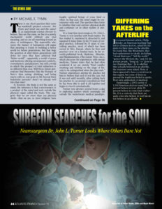
Surgeon Searches for the Soul

by Michael Tymn

Posted on

Smallpox: Curse Since Ancient Egypt?

Smallpox was once one of humanity’s most devastating diseases, but its origin is shrouded in mystery. For years, scientific estimates of when the smallpox virus first emerged have been at odds with historical records. Now, a new study reveals that the virus dates back 2,000 years further than scientists have previously shown, verifying historical sources and confirming for the first time that the disease has plagued human societies since ancient times.

The paper appears in the journal Microbial Genomics, published by the Microbiology Society.
Smallpox, caused by the variola virus, is perhaps best known for being the only infectious human disease to be eradicated worldwide. But the disease was a major cause of death until relatively recently, killing at least 300 million people in the 20th century. This is roughly the equivalent of the population of the United States.
Until relatively recently, the earliest genetic evidence for smallpox was only from the 1600s. Then in 2020, a study that sampled skeletal and dental remains of Viking-age skeletons recovered multiple strains of variola and confirmed the virus’ existence at least another 1,000 years earlier.

However, some historians believe that smallpox has been around since long before the Vikings. Suspicious scarring on ancient Egyptian mummies (including the Pharoah Ramses V who died in 1157 BC) leads some to believe that the history of smallpox stretches back at least 3,000 years. So far, the missing piece of scientific evidence to support this theory has remained hidden.

By comparing the genomes of modern and historic strains of variola virus, researchers at the Scientific Institute Eugenio Medea and University of Milan in Italy have traced the evolution of the virus back in time. They found that different strains of smallpox all descended from a single common ancestor and that a small fraction of the genetic components found in Viking-age genomes had persisted until the 18th century.

They also worked out an estimate for when the virus originated. In their estimate, the researchers accounted for something called the ‘time-dependent rate phenomenon’. This means that the speed of evolution depends on the length of time over which it is being measured, so viruses appear to change more quickly over a short timeframe and more slowly over a longer timeframe. The phenomenon has been well-documented in DNA viruses like variola.
Using a mathematical equation, scientists can account for the time-dependent rate phenomenon to give more accurate dates for evolutionary events, such as the appearance of a new virus. This gave the team a new estimate for the first emergence of smallpox: more than 3,800 years ago. Just as historians have long suspected.

The researchers hope these findings will settle a longstanding controversy and provide new insight into the history of one of humanity’s deadliest diseases.

Picture and caption:
https://microbiologysociety.org/news/press-releases/smallpox-has-plagued-humans-since-ancient-egyptian-times-new-evidence-confirms.html
https://www.microbiologyresearch.org/content/journal/mgen/10.1099/mgen.0.000932

AR #86

Mystery of the Montauk Monster

by Steven Sora