Posted on

Conscious-Like Activity Found In the Dying Brain

Reports of near-death experiences—with tales of white light, visits from departed loved ones, hearing voices, among other attributes—capture our imagination and are deeply engrained in our cultural landscape.

The fact that these reports share so many common elements begs the question of whether there is something fundamentally real underpinning them—and that those who have managed to survive death are providing glimpses of a consciousness that does not completely disappear, even after the heart stops beating.

A study published in the Proceedings of the National Academy of Science, provides early evidence of a surge of activity correlated with consciousness in the dying brain.

The study, led by Jimo Borjigin, Ph.D., associate professor in the Department of Molecular & Integrative Physiology and the Department of Neurology, and her team is a follow up to animal studies conducted almost 10 years ago in collaboration with George Mashour, M.D., Ph.D., the founding director of the Michigan Center for Consciousness Science.

Similar signatures of gamma activation were recorded in the dying brains of both animals and humans upon a loss of oxygen following cardiac arrest.

“How vivid experience can emerge from a dysfunctional brain during the process of dying is a neuroscientific paradox. Dr. Borjigin has led an important study that helps shed light on the underlying neurophysiologic mechanisms,” said Mashour.

The team identified four patients who passed away due to cardiac arrest in the hospital while under EEG monitoring. All four of the patients were comatose and unresponsive. They were ultimately determined to be beyond medical help and, with their families’ permission, removed from life support.

Upon removal of ventilator support, two of the patients showed an increase in heart rate along with a surge of gamma wave activity, considered the fastest brain activity and associated with consciousness.

Furthermore, the activity was detected in the so-called hot zone of neural correlates of consciousness in the brain, the junction between the temporal, parietal and occipital lobes in the back of the brain. This area has been correlated with dreaming, visual hallucinations in epilepsy, and altered states of consciousness in other brain studies.

These two patients had previous reports of seizures, but no seizures during the hour before their deaths, explains Nusha Mihaylova, M.D., Ph.D., a clinical associate professor in the Department of Neurology who has collaborated with Borjigin since 2015 by collecting EEG data from deceased patients under intensive care unit treatment. The other two patients did not display the same increase in heart-rate upon removal from life support, nor did they have increased gamma activity.

Because of the small sample size, the authors caution against making any global statements about the implications of the findings. They also note that it’s impossible to know in this study what the patients experienced because they did not survive.

“We are unable to make correlations of the observed neural signatures of consciousness with a corresponding experience in the same patients in this study. However, the observed findings are definitely exciting and provide a new framework for our understanding of covert consciousness in the dying humans,” she said.

Larger, multi-center studies including EEG-monitored ICU patients who survive cardiac arrest, could provide much needed data to determine whether or not these bursts in gamma activity are evidence of hidden consciousness even near death.

Additional authors on this paper include Gang Xu, Duan Li, Fangyun Tian, Peter M. Farrehi, Jack M. Parent and Michael Wang Paper cited: “Surge of neurophysiological coupling and connectivity of gamma oscillations in the dying human brain,” Proceedings of the National Academy of Science. https://www.pnas.org/doi/10.1073/pnas.2216268120

AR #106

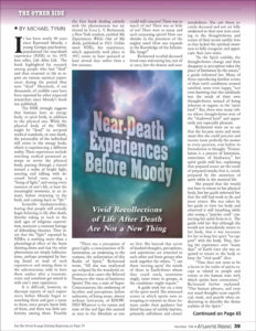
Near Death Experience Before Moody

by Michael Tymn

Posted on

Does Newly Discovered Intracellular Electricity Power Biology?

The human body relies heavily on electrical charges. Lightning-like pulses of energy fly through the brain and nerves and most biological processes depend on electrical ions traveling across the membranes of each cell in our body.

These electrical signals are possible, in part, because of an imbalance in electrical charges that exists on either side of a cellular membrane. Until recently, researchers believed the membrane was an essential component to creating this imbalance. But that thought was turned on its head when researchers at Stanford University discovered that similar imbalanced electrical charges can exist between microdroplets of water and air.

Now, researchers at Duke University have discovered that these types of electric fields also exist within and around another type of cellular structure called biological condensates. Like oil droplets floating in water, these structures exist because of differences in density. They form compartments inside the cell without needing the physical boundary of a membrane.

Inspired by previous research demonstrating that microdroplets of water interacting with air or solid surfaces create tiny electrical imbalances, the researchers decided to see if the same was true for small biological condensates. They also wanted to see if these imbalances sparked reactive oxygen,  “redox,” reactions like these other systems.
“In a prebiotic environment without enzymes to catalyze reactions, where would the energy come from? This discovery provides a plausible explanation of where the reaction energy could have come from, just as the potential energy that is imparted on a point charge placed in an electric field.” (https://www.sciencedirect.com/science/article/abs/pii/S2451929423001535)

Because the Chilkoti laboratory specializes in creating synthetic versions of naturally occurring biological condensates, the researchers were easily able to create a test bed for their theory. After combining the right formula of building blocks to create minuscule condensates, with help from postdoctoral scholar Marco Messina in? Christopher J. Chang’s group at the University of California – Berkeley, they added a dye to the system that glows in the presence of reactive oxygen species.

Their hunch was right. When the environmental conditions were right, a solid glow started from the edges of the condensates, confirming that a previously unknown phenomenon was at work. Dai next talked with Richard Zare, the Marguerite Blake Wilbur Professor of Chemistry at Stanford, whose group established the electric behavior of water droplets. Zare was excited to hear about the new behavior in biological systems, and started to work with the group on the underlying mechanism.

AR #38

Rupert Sheldrake’s Seven Senses

by Cynthia Logan

Posted on

Brushing with Shapeshifting Microrobots

A shapeshifting robotic microswarm may one day act as a toothbrush, rinse, and dental floss in one.

The technology, developed by a multidisciplinary team at the University of Pennsylvania, is poised to offer a new and automated way to perform the mundane but critical daily tasks of brushing and flossing. It’s a system that could be particularly valuable for those who lack the manual dexterity to clean their teeth effectively themselves.
The building blocks of these microrobots are iron oxide nanoparticles that have both catalytic and magnetic activity. Using a magnetic field, researchers could direct their motion and configuration to form either bristle-like structures that sweep away dental plaque from the broad surfaces of teeth, or elongated strings that can slip between teeth like a length of floss. In both instances, a catalytic reaction drives the nanoparticles to produce antimicrobials that kill harmful oral bacteria on site.


Experiments using this system on mock and real human teeth showed that the robotic assemblies can conform to a variety of shapes to nearly eliminate the sticky biofilms that lead to cavities and gum disease. The Penn team shared their findings establishing a proof-of-concept for the robotic system in the journal ACS Nano.


“Routine oral care is cumbersome and can pose challenges for many people, especially those who have hard time cleaning their teeth” says Hyun (Michel) Koo, a professor in the Department of Orthodontics and divisions of Community Oral Health and Pediatric Dentistry in Penn’s School of Dental Medicine and co-corresponding author on the study. “You have to brush your teeth, then floss your teeth, then rinse your mouth; it’s a manual, multistep process. The big innovation here is that the robotics system can do all three in a single, hands-free, automated way.”


“Nanoparticles can be shaped and controlled with magnetic fields in surprising ways,” says Edward Steager, a senior research investigator in Penn’s School of Engineering and Applied Science and co-corresponding author. “We form bristles that can extend, sweep, and even transfer back and forth across a space, much like flossing. The way it works is similar to how a robotic arm might reach out and clean a surface. The system can be programmed to do the nanoparticle assembly and motion control automatically.”


Hyun (Michel) Koo is a professor in the Department of Orthodontics and divisions of Community Oral Health and Pediatric Dentistry in the School of Dental Medicine and co-director of the Center for Innovation & Precision Dentistry at the University of Pennsylvania.


Edward Steager is a senior research investigator in Penn’s School of Engineering and Applied Science.
Koo and Steager’s coauthors on the paper are Penn Dental Medicine’s Min Jun Oh, Alaa Babeer, Yuan Liu, and Zhi Ren and Penn Engineering’s Jingyu Wu, David A. Issadore, Kathleen J. Stebe, and Daeyeon Lee.


This work was supported in part by the National Institute for Dental and Craniofacial Research (grants DE025848 and DE029985), Procter & Gamble, and the Postdoctoral Research Program of Sungkyunkwan University.

https://penntoday.upenn.edu/news/penn-dental-engineering-shapeshifting-microrobots-can-brush-and-floss-teeth

Atlantis Rising Issue #58
9,000-Year-Old Dental Drilling

Early Rays


Posted on

Spirituality vs. Heart Failure

Numerous studies have shown that spirituality can help improve quality of life for people with chronic diseases like cancer. According to a literature review published today in JACC: Heart Failure, spirituality can also have a positive impact on quality of life for heart failure patients. It further concludes spirituality should be considered a potential target for palliative care interventions to improve patient-centered and clinical outcomes in these individuals.

“Patients who have heart failure experience a poorer quality of life compared to their peers, with high levels of depression, anxiety and spiritual distress,” said Rachel S. Tobin, MD, resident in Internal Medicine at Duke University Hospital, and lead author of the study. “Contributing to diminished quality of life is the fact that heart failure, unlike many other chronic diseases, is very unpredictable and can lead to hopelessness, isolation and altered self-image.”
The American College of Cardiology and other major cardiovascular societies recommend palliative care for heart failure patients. Spirituality is a core domain of palliative care, with the goal of identifying and addressing spiritual concerns and providing patients with appropriate spiritual and religious resources. However, limited research has been conducted on spirituality’s impact on patients with heart failure, and there are no known tools designed to measure it.


According to the researchers, spirituality is hard to define, but they reference several definitions that describe spirituality as how individuals find meaning and purpose in life, which can be separate from religious beliefs.
For instance, the Institute of Medicine defines spirituality as “the needs and expectations which humans have to find meaning, purpose and value in their life. Such needs can be specifically religious, but even people who have no religious faith or are not members of an organized religion have belief systems that give their lives meaning and purpose.”

https://www.acc.org/About-ACC/Press-Releases/2022/03/28/18/13/Spirituality-Can-Improve-Quality-of-Life-for-Heart-Failure-Patients

Atlantis Rising Issue #81
The Doomsday Fallacy

by John White

Posted on

Is Heart Fabrication Getting Closer?

Heart disease — the leading cause of death in the U.S. — is so deadly in part because the heart, unlike other organs, cannot repair itself after injury. That is why tissue engineering, ultimately including the wholesale fabrication of an entire human heart for transplant, is so important for the future of cardiac medicine. 

To build a human heart from the ground up, researchers need to replicate the unique structures that make up the heart. This includes recreating helical geometries, which create a twisting motion as the heart beats. It’s been long theorized that this twisting motion is critical for pumping blood at high volumes, but proving that has been difficult, in part because creating hearts with different geometries and alignments has been challenging.


Now, bioengineers from the Harvard John A. Paulson School of Engineering and Applied Sciences (SEAS) have developed the first biohybrid model of human ventricles with helically aligned beating cardiac cells, and have shown that muscle alignment does, in fact, dramatically increases how much blood the ventricle can pump with each contraction.


This advancement was made possible using a new method of additive textile manufacturing, Focused Rotary Jet Spinning (FRJS), which enabled the high-throughput fabrication of helically aligned fibers with diameters ranging from several micrometers to hundreds of nanometers. Developed at SEAS by Kit Parker’s Disease Biophysics Group, FRJS fibers direct cell alignment, allowing for the formation of controlled tissue engineered structures.
The research is published in Science. 


“This work is a major step forward for organ biofabrication and brings us closer to our ultimate goal of building a human heart for transplant,” said Parker, the Tarr Family Professor of Bioengineering and Applied Physics at SEAS and senior author of the paper.

https://www.seas.harvard.edu/news/2022/07/major-step-forward-organ-biofabrication

AR #79

Surgeon Searches for the Soul

by Michael Tymn


Posted on

Could Goji Berries Help Seniors to See the light

Regularly eating a small serving of dried goji berries may help prevent or delay the development of age-related macular degeneration, or AMD, in healthy middle-aged people, according to a small, randomized trial conducted at the University of California, Davis.

AMD is the leading cause of vision loss in older people, and is estimated to affect more than 11 million in the United States and 170 million globally. 


“AMD affects your central field of vision and can affect your ability to read or recognize faces,” said Glenn Yiu, a co-author of the study and an associate professor in the Department of Ophthalmology and Vision Sciences.
The researchers found that 13 healthy participants aged 45 to 65 who consumed 28 grams (about one ounce, or a handful) of goji berries five times a week for 90 days increased the density of protective pigments in their eyes. In contrast, 14 study participants who consumed a commercial supplement for eye health over the same period did not show an increase.


The pigments that increased in the group that ate goji berries, lutein and zeaxanthin, filter out harmful blue light and provide antioxidant protection. Both help to protect the eyes during aging. 
“Lutein and zeaxanthin are like sunscreen for your eyes,” said lead author Xiang Li, a doctoral candidate in the Nutritional Biology Program.


“The higher the lutein and zeaxanthin in your retina, the more protection you have. Our study found that even in normal healthy eyes, these optical pigments can be increased with a small daily serving of goji berries,” said Li.
The study was published in the journal Nutrients.


Goji berries are the fruit of Lycium chinense and Lycium barbarum, two species of shrubby bushes found in northwest China. The dried berries are a common ingredient in Chinese soups and are popular as herbal tea. They are similar to raisins and eaten as a snack.


In Chinese medicine, goji berries are said to have “eye brightening” qualities. Li grew up in northern China and became curious whether there were any physiological properties to “eye brightening.”


“Many types of eye diseases exist, so it is not clear which disease ‘eye brightening’ is targeting,” said Li.
She researched the bioactive compounds in goji berries and found they contain high quantities of lutein and zeaxanthin, which are known to reduce the risk of eye diseases related to AMD. The form of zeaxanthin in goji berries is also a highly bioavailable form, according to Li, meaning it is readily absorbed in the digestive system so the body can use it.  


The current treatment for intermediate stages of AMD uses special dietary supplements, called AREDS, that contain vitamins C, E, zinc, copper and lutein and zeaxanthin. No known therapy has yet been shown to impact early stages of AMD.


The cause of AMD is complex and multifactorial, according to Yiu, and involves a mix of genetic risks, age-associated changes, and environmental factors like smoking, diet and sun exposure. Early stages of AMD do not have symptoms; however, physicians can detect AMD and other eye problems during a regular comprehensive eye exam.
“Our study shows goji berries, which are a natural food source, can improve macular pigments of healthy participants beyond taking high-dose nutritional supplements,” said Yiu. “The next step for our research will be to examine goji berries in patients with early-stage AMD.”


Although the results are promising, the researchers note that the study size was small and more research will be needed.

https://www.ucdavis.edu/health/news/dried-goji-berries-may-provide-protection-against-age-related-vision-loss

AR #132

When Color Vision Becomes a Super Power

Alternative News


Posted on

Could Breastfeeding Fight Cognitive Decline in Moms?

A new study led by researchers at UCLA Health has found that women over the age of 50 who had breastfed their babies performed better on cognitive tests compared to women who had never breastfed. The findings, published in Evolution, Medicine and Public Health, suggest that breastfeeding may have a positive impact on postmenopausal women’s cognitive performance and could have long-term benefits for the mother’s brain.

“While many studies have found that breastfeeding improves a child’s long-term health and well-being, our study is one of very few that has looked at the long-term health effects for women who had breastfed their babies,” said Molly Fox, PhD, lead author of the study and an Assistant Professor in the UCLA Department of Anthropology and the Department of Psychiatry and Biobehavioral Sciences. “Our findings, which show superior cognitive performance among women over 50 who had breastfed, suggest that breastfeeding may be ‘neuroprotective’ later in life.”
Cognitive health is critical for wellbeing in aging adults. Yet, when cognition becomes impaired after the age of 50, it can be a strong predictor of Alzheimer’s Disease (AD), the leading form of dementia and cause of disability among the elderly – with women comprising nearly two-thirds of Americans living with the disease.


Many studies also show that phases of a woman’s reproductive life-history, such as menstruation, pregnancy, breastfeeding and menopause can be linked to a higher or lower risk for developing various health conditions like depression or breast cancer, yet few studies have examined breastfeeding and its impact on women’s long-term cognition. Of those that have, there has been conflicting evidence as to whether breastfeeding might be linked to better cognitive performance or Alzheimer’s risk among post-menopausal women.


“What we do know is that there is a positive correlation between breastfeeding and a lower risk of other diseases such as type-2 diabetes and heart disease, and that these conditions are strongly connected to a higher risk for AD,” said Helen Lavretsky, MD, the senior author of the study and a professor in the Department of Psychiatry and Biobehavioral Sciences at the Semel Institute for Neuroscience and Human Behavior at UCLA. 


“Because breastfeeding has also been found to help regulate stress, promote infant bonding and lower the risk of post-partum depression, which suggest acute neurocognitive benefits for the mother, we suspected that it could also be associated with long-term superior cognitive performance for the mother as well,” added Dr. Fox.


To find out, the researchers analyzed data collected from women participating in two cross-sectional randomized controlled 12-week clinical trials at UCLA Health: 1) The “Brain Connectivity and Response to Tai Chi in Geriatric Depression and Cognitive Decline,” included depressed participants. 2) The “Reducing Risk for Alzheimer’s Disease in High-Risk Women through Yoga or Memory Training that included non-depressed participants with some subjective memory complaints and a risk for heart disease.


Among the two trials, 115 women chose to participate, with 64 identified as depressed and 51 non-depressed.  All participants completed a comprehensive battery of psychological tests measuring learning, delayed recall, executive functioning and processing speed. They also answered a questionnaire about their reproductive life-history that included questions about the age they began menstruating, number of complete and incomplete pregnancies, the length of time they breastfed for each child and their age of menopause.


Importantly, none of the participants had been diagnosed with dementia, or other psychiatric diagnoses such as bipolar disorder, alcohol or drug dependence, neurological disorders or had other disabilities preventing their participation or taking any psychoactive medications. There was also no significant difference in age, race, education or other cognitive measures between the depressed and non-depressed participants.


Key findings from the researchers’ analysis of the data collected from questionnaires on the women’s reproductive history revealed that about 65% of non-depressed women reported having breastfed, compared to 44% of the depressed women. All non-depressed participants reported at least one completed pregnancy compared to 57.8% of the depressed participants.


Results from the cognitive tests also revealed that those who had breastfed, regardless of whether they were depressed or not, performed better in all four of the cognitive tests measuring for learning, delayed recall, executive functioning and processing compared to women who had not breastfed.


AR #105

Morphic Fields & DNA
by William B. Stoecker